Oncological surgery removes what cannot remain. What follows the reconstruction must be as precise as the resection. We engineer for both.
Oncological surgery presents a reconstruction challenge unlike any other: resection margins, remaining bone stock, compromised soft tissue, and prior treatment each shape a post-resection anatomy that no off-the-shelf implant was designed for. Every defect is singular.
Atomic Additive works with oncological surgical teams across neurosurgery, orthopaedics, and CMF to provide patient-specific reconstruction implants designed from the actual post-resection defect — planned in collaboration with the surgical team before the procedure, so that reconstruction begins the moment resection ends.
For oncological cases, preparation is not a convenience. It is a clinical necessity.
Seven products drawn across neurosurgery, orthopaedics, and CMF — each designed around the specific post-resection defect.
Custom cranial and spinal implants for reconstruction following tumour resection — designed around the specific defect geometry post-excision, engineered to restore structural integrity.
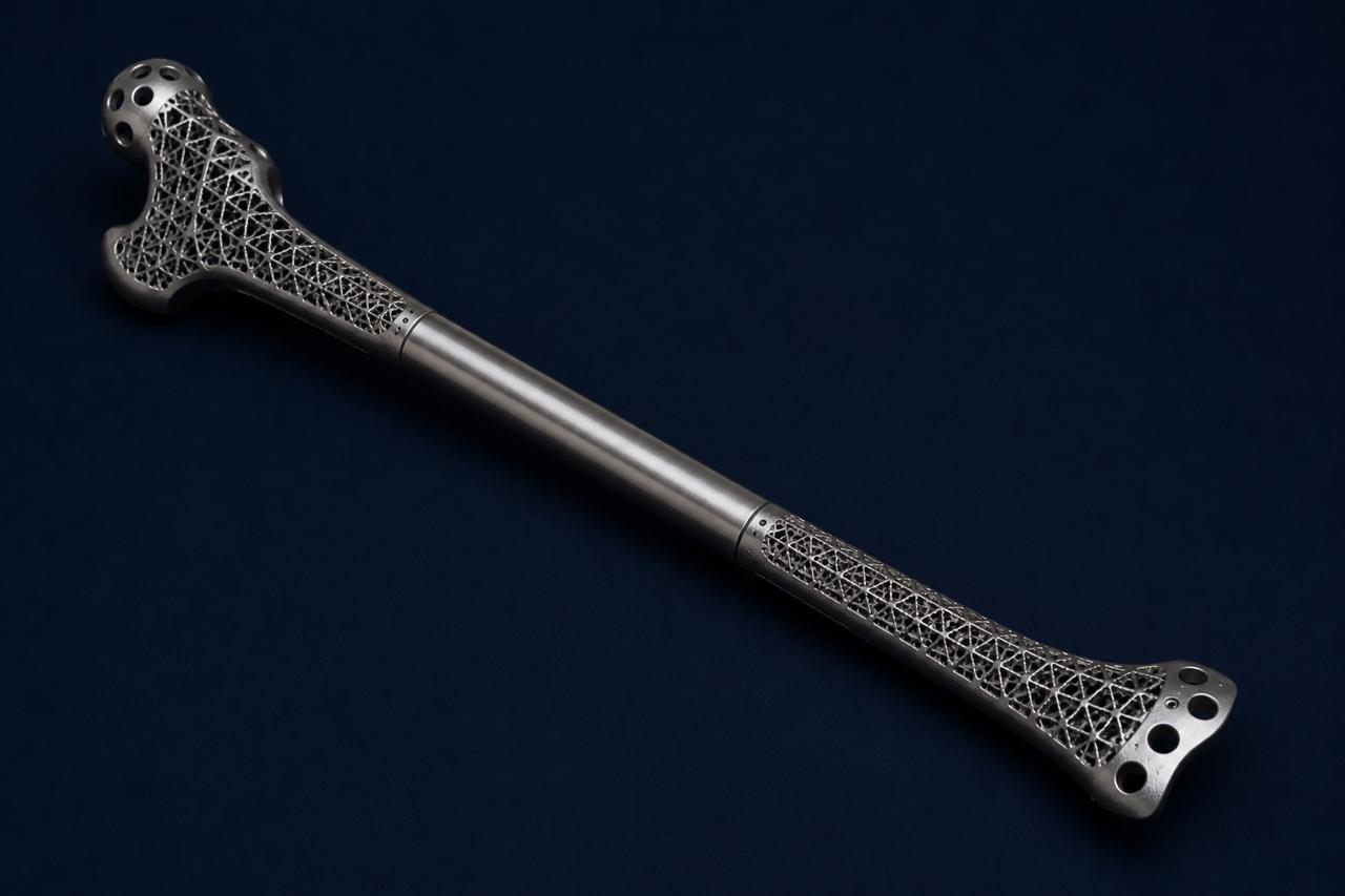
Patient-specific implants for long bone and appendicular skeletal reconstruction following primary bone tumour resection — restoring structural continuity and enabling functional recovery.
Custom pelvic implants for post-oncological hemipelvectomy and internal pelvic resection — one of the most demanding reconstruction challenges in surgical oncology, requiring precise fit and load transfer.
Custom prosthetic systems for limb-preserving surgery in bone and soft tissue sarcoma — engineered to maximise function and structural durability post-resection, preserving the limb while achieving oncological control.
Patient-matched plates for jaw reconstruction following oncological mandibular resection — restoring function, occlusion, and facial form in post-resection reconstruction.
Patient-specific models of tumour and surrounding anatomy for pre-resection planning, margin definition, and multidisciplinary team briefing — enabling consensus before the patient enters theatre.
Patient-matched cutting guides that translate planned resection margins to the surgical field — ensuring oncologically safe and geometrically accurate bone cuts that match the preoperative plan precisely.
3D tumour mapping and post-resection defect modelling from DICOM data — enabling implant design to begin before surgery and reconstruction to start the moment resection ends.
Learn more →Custom oncological reconstruction implants — cranial, spinal, pelvic, long bone, and maxillofacial — engineered around the specific defect geometry and surgeon-reviewed before manufacture.
Learn more →Custom cutting guides for oncological bone resection — translating planned margins to the surgical field to achieve wide, clear margins while preserving adjacent neurovascular structures.
Learn more →Physical tumour models for MDT briefing, margin planning, and surgical rehearsal — enabling the entire team to understand the anatomy and agree on approach before the first incision.
Learn more →Brain, spine, and skull — where anatomy is most complex, space is most constrained, and the margin for error is smallest.
Learn more → SPECIALITY 02Complex reconstruction, revision arthroplasty, and limb salvage — for the cases where standard catalogues reach their limit.
Learn more → SPECIALITY 03Restoring form and function with anatomical precision — millimetre accuracy for orbital, mandibular, and midface reconstruction.
Learn more →Submit your case and our clinical team will be in touch within 24 hours — we will discuss your resection plan, defect geometry, and how best to prepare the reconstruction before surgery begins.